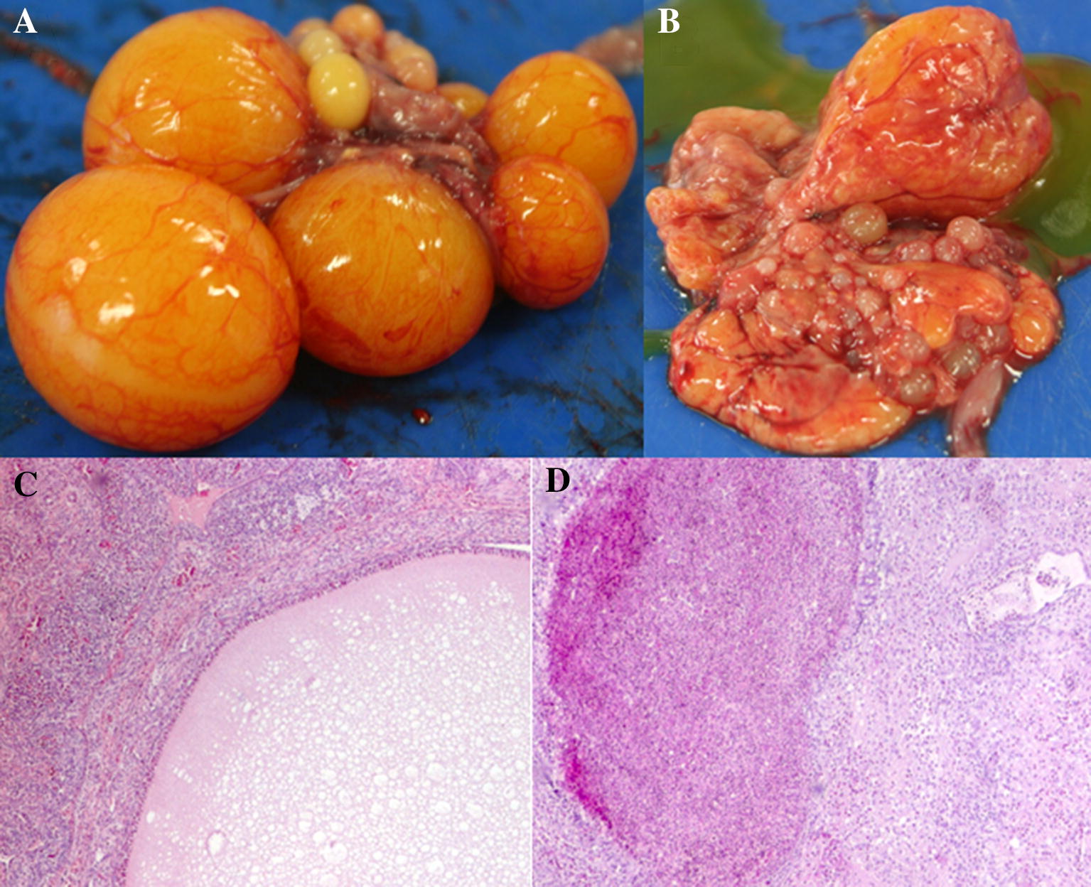
Figure 1.

Gross lesions, histopathology and apoptotic cells found in chickens infected withG. anatisΔgtxAmutant orG. anatiswild-type (WT) strain. Ovaries from 29-week old laying hens. Two days post-inoculation with either G. anatis ΔgtxA or G. anatis WT. AG. anatis ΔgtxA infection group. Oophoritis with vascular engorgement and slight edema. BG. anatis wild-type. Diffuse purulent oophoritis and folliculitis with ruptures follicles. C Hematoxylin and eosin (HE)-straining of the follicle in ovarian tissue from the G. anatis ΔgtxA infection group. Focal oophoritis with the presence of inflammatory cells in the stroma and vascular engorgement. Infiltrates of few heterophilic granulocytes and mononuclear cells. D HE-straining of the ovary of the G. anatis WT infection group. Purulent oophoritis with heavy infiltration of inflammatory cells and granuloma formation (black arrows) in the follicle.
