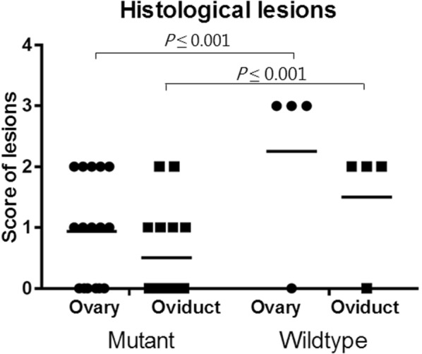
Figure 2.

Score of histological lesions in the ovary and oviduct of chickens infected withG. anatisΔgtxAorG. anatiswild-type strain. No clinical signs of infection were observed after inoculation. At the necropsy, 10 out of the 16 birds having received G. anatis ΔgtxA demonstrated mild lesions, whereas remaining birds did not have any lesions. In birds infected with G. anatis WT strain, three of four birds had severe lesions and one was without lesions. No lesions were found in the control bird group. The number of organs with lesions in birds from the ΔgtxA group was significantly lower compared to the number of organs with lesions in WT group (P = 0.003). No difference was found between a number of lesions when comparing birds examined at 2 or 6 days pi from the ΔgtxA group (P = 0.2). In the ΔgtxA infection group, a mild non-purulent oophoritis characterized by vascular congestion and enlargement of the stromal tissue of the ovary (Figure 1A) was observed in five birds. In three birds, the oophoritis was purulent and in two birds, both examined 6 days pi, focal purulent oophoritis and localized peritonitis was found. Salpingitis was not observed in any of the birds from the ΔgtxA mutant infection group. The three out of four birds in the WT strain group had gross lesions, all exhibiting diffuse purulent peritonitis, diffuse purulent oophoritis and salpingitis (Figure 1B). No lesions were found in the spleen or liver of any birds. * indicates a statistically significant difference between groups (P ≤ 0.05).
