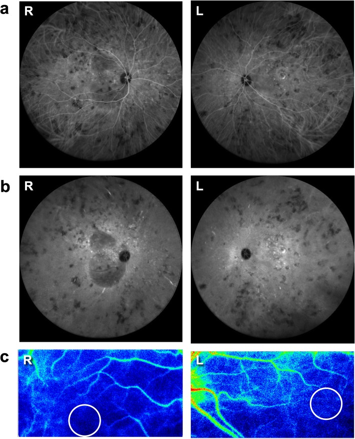
Fig. 2.
Indocyanine green angiography (ICGA) and laser speckle flowgraphy (LSFG) at the first visit. ICGA detected fuzzy vascular pattern of large stromal vessels in the mid-venous phase (a) and sharply marginated hypofluorescent lesions of various sizes that were clearly observed throughout the mid-venous phase (a) and the late phase (b). LSFG showed a cold-color pattern of the color map in the macular area bilaterally, and the mean blur rate (MBR) values in the circle were 2.16 in the right eye (R) and 2.63 in the left eye (L) (c). R: right, L: left

