Figure 2.

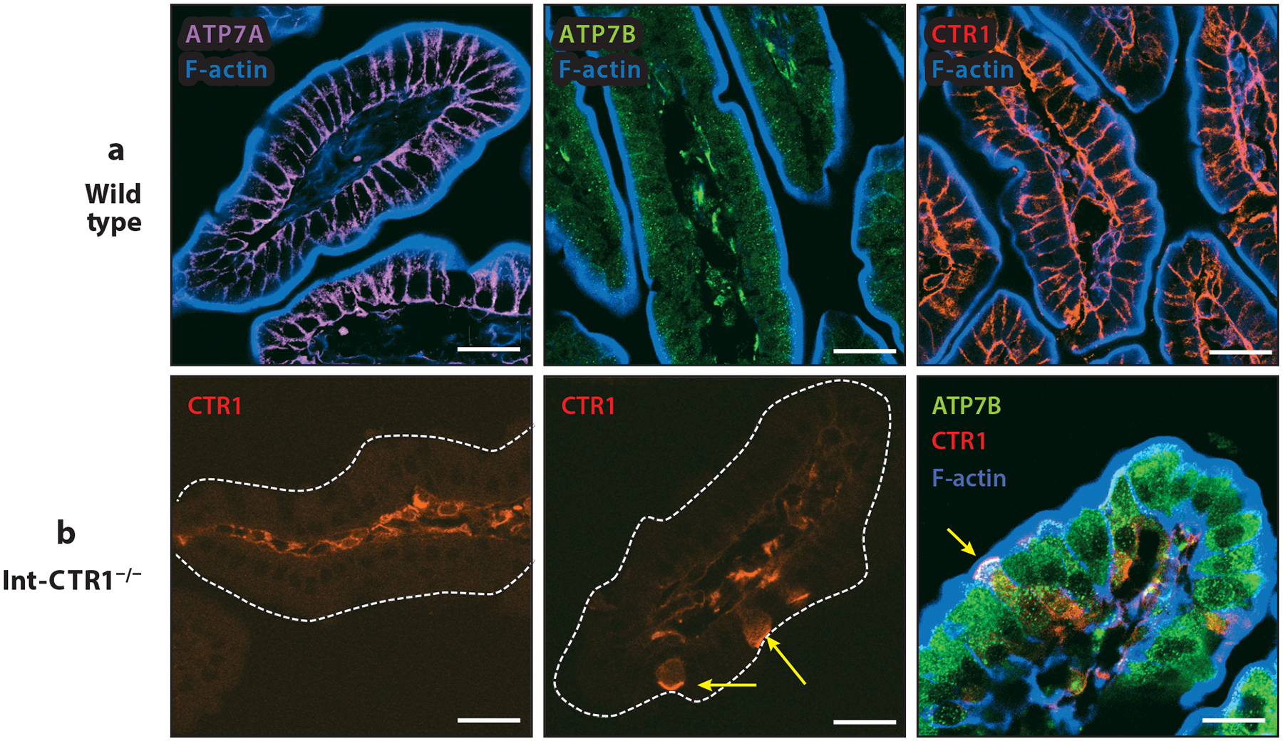
Expression and distribution of ATP7A, ATP7B, and CTR1 in murine small intestine. Duodenal intestinal tissues from 2-week-old sex-matched mice were used to assess the expression and intracellular distribution of the key players in intestinal copper (Cu) homeostasis. (a) In wild-type tissues, ATP7A (probed with anti-ATP7A sc376467, Santa Cruz Biotechnology) showed strong basolateral staining consistent with previous reports. ATP7B (probed with anti-ATP7b ab124973, Abcam) had a strong vesicular pattern, and CTR1 (probed with 8G11-F10, hybridoma monoclonal antibody; H. Pierson, H. Yang & S. Lutsenko, unpublished data) was detected specifically at the basolateral membrane. F-actin (blue) marks the apical cell border. (b) Tissues with intestine-specific deletion of CTR1 in enterocytes (Int-CTR1−/−) specifically lack CTR1 expression in the intestinal epithelium and show no basolateral staining of CTR1 in the epithelial layer. However, sparse epithelial cells did show CTR1 expression in the apical membrane in the context of the Int-CTR1−/− tissues (yellow arrows). All scale bars: 25 μm.
