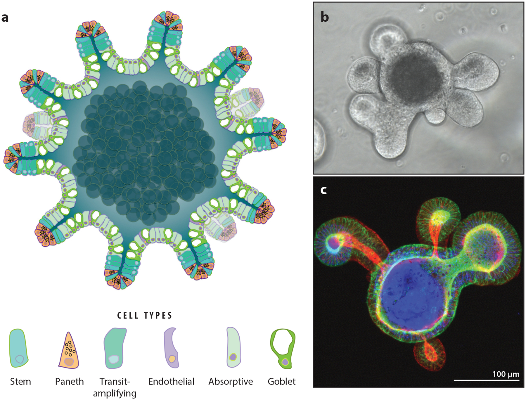
Figure 3.

Three-dimensional (3D) enteroids: sophisticated model systems for investigating intestinal epithelium. (a) Schematic of an enteroid. Small crypt-like projections decorate the surface of the enteroid. The apical cell surface faces the lumen, analogous to the morphology of the digestive tract. Panel a adapted with permission from Reference 84. (b) Bright-field image of a single, live differentiated mouse enteroid in culture. (c) 3D projection of a single differentiated mouse enteroid with the apical membranes stained for F-actin demonstrates apical–inward polarity.
