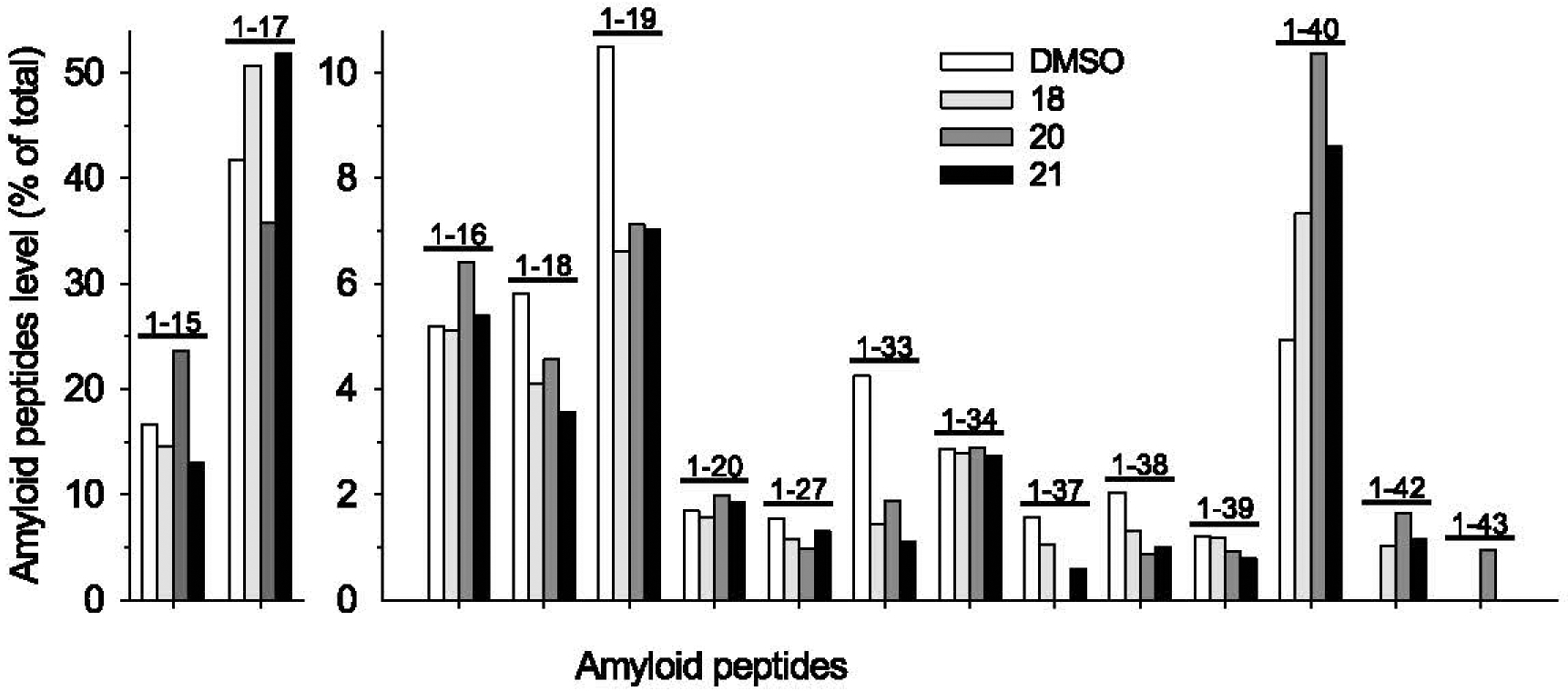
Fig. 6.

Pattern of Aβs produced by N2a-APP695 cells exposed to pyrazoles 18, 20, or 21. Cells were treated for 18 h with DMSO or 20 μM of each pyrazole. Cell supernatants were collected and analyzed as described. Quantification of all Aβs in N2a-APP695 cells supernatants are presented as percentage of total amyloids. Note the decrease in peptides Aβ1–19, Aβ1–37, Aβ1–38, the increase in Aβ1–40 and the appearance of Aβ1–42 and Aβ1–43 in the supernatants of pyrazole-treated cells (these two peptides are undetectable in the supernatant of DMSO-treated cells).
