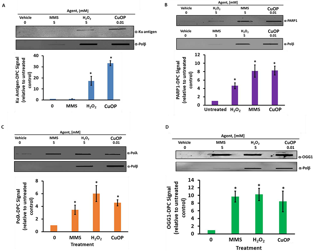
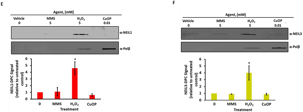
Figure 3. Chemical specificity of AP Lyase-DPC formation.


MDA-MB-231 cells were exposed for 30 min to 5 mM MMS or 5 mM H2O2, or to 10 μM CuOP for 1 h (under conditions described in section 2.1), and gDNA was immediately isolated. Samples (1 μg) were assayed for DPC containing: A, Ku antigen; B, PARP1; C, Polλ; D, OGG1; E, NEIL1; F, NEIL3. For each set of experiments, a representative blot is shown, with quantification for n= 3 independent experiments, normalized to untreated cells. Error bars show +/−S.D. * indicates p<0.05 (two-tailed Student’s t-test) compared to untreated control.
