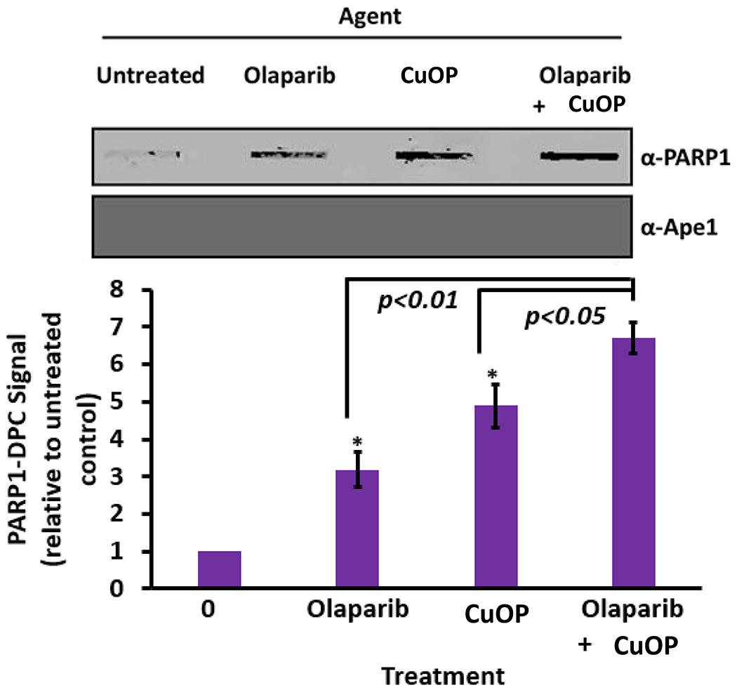
Figure 4. Effect of PARP inhibitor on oxidative PARP1-DPC formation.

MDA-MB-231 cells were exposed for 1.5 h to olaparib or CuOP, or to both sequentially (under conditions described in section 2.1). For the combination treatment, the cells were first exposed to 10 μM CuOP for 1 h, followed by addition of 10 μM olaparib for 30 min. Immediately after the treatments, gDNA was isolated. Aliquots of gDNA (1 μg) were assayed for PARP1-DPC; blots were also probed for Ape1 as a negative control. Representative blots are shown, with quantification for n= 3 independent experiments, normalized to untreated cells. Error bars show +/−S.D. for n= 3 independent experiments. * indicates p<0.05 or p<0.01 (two-tailed Student’s t-test) compared to the indicated treated controls.
