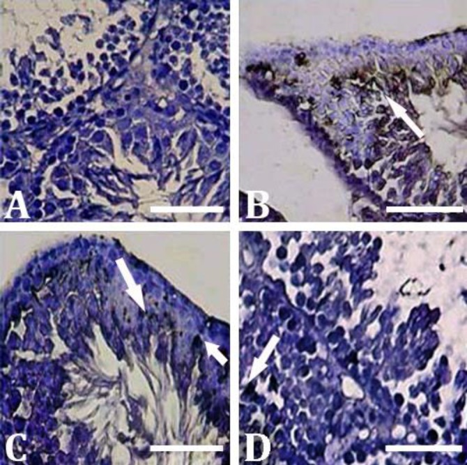
Fig. 3.

Histochemical analyses for the presence of ALP in testicular tissue. A) Control group; B) MTX group, intensive increase in ALP activity (arrows); C) MTX/CMFE/500 group; and D) MTX/CMFE/1000 group. ALP activity was significantly decreased (p < 0.05) in CMFE treated animals (Alkaline phosphatase staining, Bar = 60 µm)
