Figure 2.

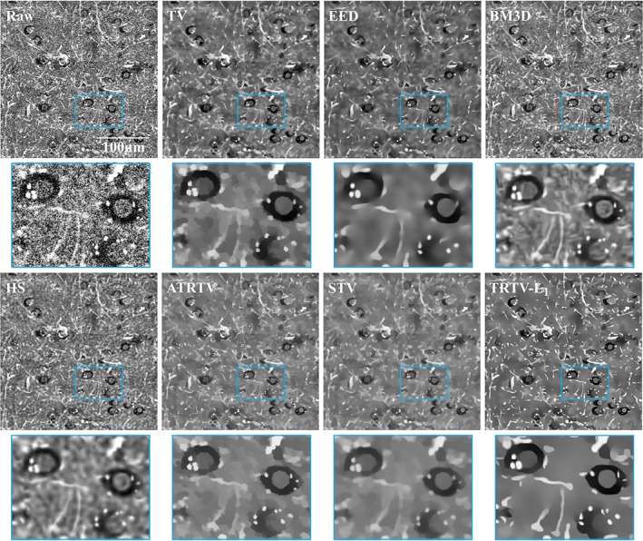
One 2D THG image of normal brain tissue from gray matter. Brain cells and neuropil appear as dark holes with dimly seen nuclei inside and bright fibers, respectively

One 2D THG image of normal brain tissue from gray matter. Brain cells and neuropil appear as dark holes with dimly seen nuclei inside and bright fibers, respectively