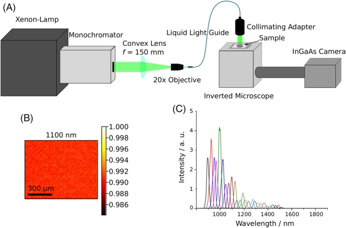
Figure 2.

Schematic diagram of the multispectral NIR absorption setup. A, General overview of the setup consisting of a broadband light source, a monochromator, optics to enable wide‐field transmission illumination, an inverted microscope and a cooled NIR InGaAs camera. B, Image acquired with the monochromator setup on a blank sample, intensity normalized to the highest intensity in the image. The high homogeneity of the intensity distribution in the image is clearly observable. C, Spectra of the monochromator lamp. The FWHM of the single Gaussians are about 25 nm and the setup covered potentially the spectral range from 400 nm up to 1500 nm. For this experiment, the range between 900 and 1500 nm only was used
