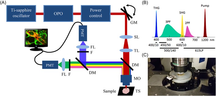
Figure 1.

HHG microscopy for breast tissue imaging. A, Multi‐photon microscope setup: A laser source produces pulses of 200 fs at 1200 nm. The laser beam passes a pair of galvo‐scanner mirrors (GM), scan lens (SL), tube lens (TL) and is focused into the sample with a microscope objective (MO). The first dichroic mirror reflects backscattered HHG signals, and a second one splits them into a first channel (THG) and a second (SHG/2PF/3PF) channel. The signals are filtered by interference filters (F) and focused on the photomultiplier tube detectors (PMT) with lenses (FL). Sample can be moved with a motorized translation stage (TS). B, The light spectrum of the incoming laser beam and generated signals by the tissue, and types of filters used (indicated with black lines) to detect these signals. C, Photograph of a breast sample embedded in agar under a 0.17 mm glass cover slip in the middle of a plastic disk and the microscope objective on top
