
Figure 4.
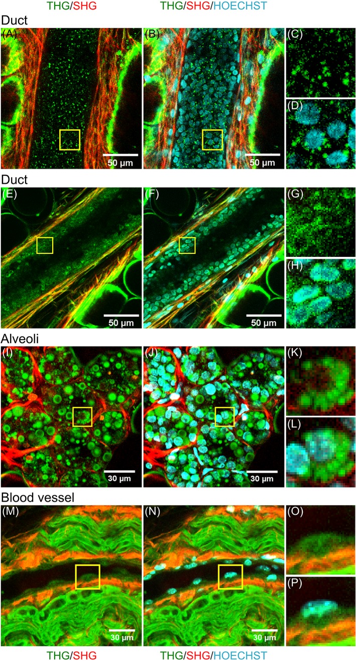
Cell nuclei in mouse breast tissue visualized with the DNA dye Hoechst‐33342. For two breast ducts of a non‐lactating mouse (A‐D, E‐H), and a lobule (I‐L) and a blood vessel surrounded by a peripheral nerve of a lactating mouse (M‐P) the combined THG (green) and SHG (red) images are shown (left), and the THG/SHG images combined with the 3PF signals from the DNA dye revealing the cell nuclei (middle). Magnified images of cells (marked with yellow squares in left and middle images) show that in the THG image the black holes in the epithelial cells of alveoli and ducts, and the thicker parts of the endothelial cells in the blood vessel contain DNA