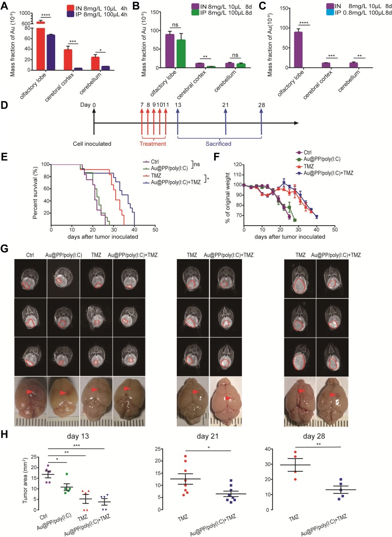
Figure 3.
Intranasal administration of Au@PP/poly(I:C) inhibited intracranial glioma growth and prolonged mice survival. (A) Accumulation of AuNPs in different brain regions in the initial stage of dosing at 8 mg/L and the amounts of Au in the brain post-injection at 4 h after IN or IP administration. (B, C) Mice were treated with 8 mg/L AuNPs every other day for 4 times by the IN and IP routes (B), while were treated with 8 mg/L AuNPs every other day for 4 times by the IN route and with 0.8 mg/L AuNPs every other day for 4 times by the IP route or treated by the IN route with a total of 10 μL of solution and by the IP route with a total of 100 μL of solution every time (C). The brain tissue was ablated with aqua regia and ICP-MS was used to detect the solution gold ion concentration. (Mean±SEM, n=6 in each group. ns: no significant difference, *p < 0.05, **p < 0.01, ***p < 0.001 and ****p < 0.0001). (D) Schematic of the therapy regimen. Intracranial glioma orthotopic xenografts were established by inoculation of GL261 cells into C57BL/6 mice. The mice received intranasal Au@PP/poly(I:C) or intragastrical TMZ every day for 5 successive days. (E) Survival of the animals. Log-rank analysis was performed for each group; for the indicated comparisons in the last two groups, ns: non-significant, *p < 0.05. (F) Body weight changes in mice after tumor inoculation. (G) Representative MRI images of the existing brain tumors in day 13, day 21 and day 28. The red circles and arrows indicate the tumors. (H) Tumor area was measured with RadiAnt DICOM Viewer. (Mean±SEM, for (A–C), n=6 in each group; for (D–F), n≥12 in each group; for G and H, n≥4. ns: no significant difference, *p < 0.05, **p < 0.01, ***p < 0.001).

