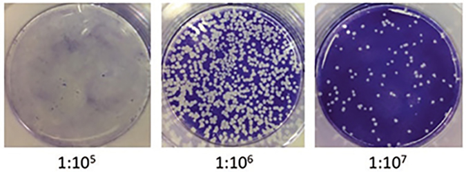
Fig. 1.
Plaque assay of VSV-infected BHK-21. BHK-21 cells were plated in a 6-well plate and left to attach overnight. The following day, serial dilutions of virus were prepared (10−5 through 10−10) and 100 μl added to the monolayer of BHK cells. After 30′ adsorption, the medium was swapped out for methylcellulose and let to solidify. The plates were then incubated at 37 °C for about 2 days. The overlay was then removed, and the cells were stained with 1% crystal violet for 20 min. The number of plaques was then counted after gently washing off the excess stain. The total number of plaque-forming units (PFU) was determined by dividing the average number of plaques by the dilution factor times the volume added

