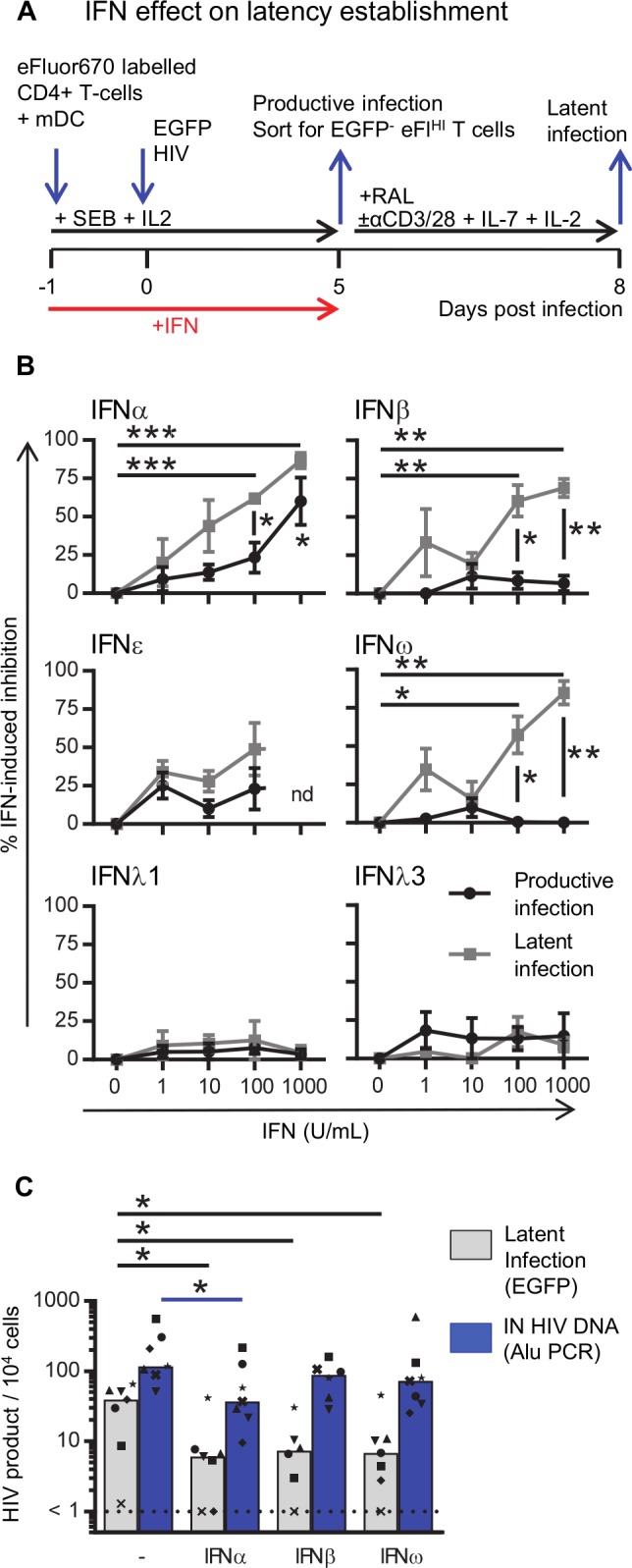
Fig 2. IFN-induced inhibition of productive infection and establishment of latent infection.

A: Experimental design to determine the effect of IFN on productive and latent infection. B: Resting CD4+ T cells were co-cultured with mDC in the absence or presence of IFN (1–1000 U/mL, as indicated) and the IFN-induced inhibition was calculated for productive (black circles) and latent (grey squares) infection. C: Resting CD4+ T cells were co-cultured with mDC in the absence and presence of 100 U/mL IFNα, IFNβ or IFNω. Cells were infected and sorted as described in A except that part of the sorted EGFP-eFluorHI population was lysed, DNA extracted and the number of HIV integrated copies was quantified using the Alu PCR and normalized to the housekeeping gene CCR5. The remainder of the sorted cells were cultured to quantify latent infection. Lines (B) indicate mean values ±SEM (n = 3–4). Columns (C) represent median values and dots represent individual donors with each donor represented by a specific symbol (n = 6–7 donors). *p<0.05, **p<0.01, ***p<0.001, as determined by paired student T test (n = 3–4) or Wilcoxon matched pairs signed rank test (n = 6–7), nd = not done.
