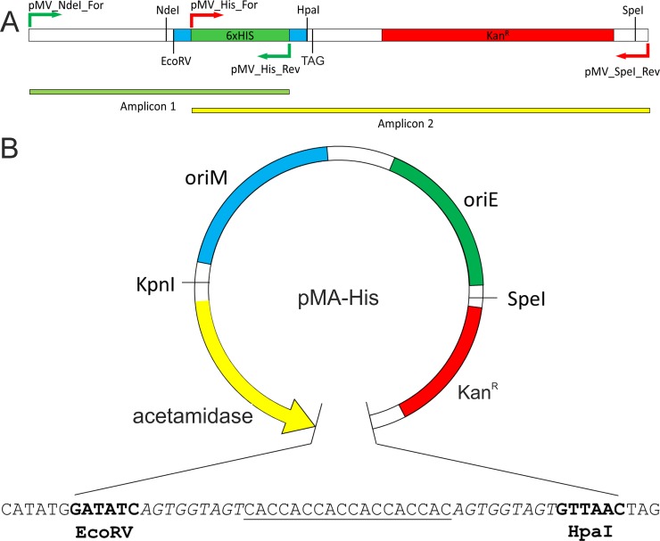
Fig 1. Schematic representation of proposed vector with pMA-His as example.
Panel A depicts the PCR-generated amplicons of 6x-His fragment (amplicon 1; green bar) and the kanamycin resistance marker (amplicon 2; yellow bar). Primers used in these PCR reactions are shown as arrows. TAG (stop codon) and relevant restriction enzyme sites are shown. Panel B shows the graphical representation of proposed plasmid with acetamide-inducible promoter system (labelled as acetamidase). oriM and oriE represent the origin of replication for mycobacteria and E. coli, respectively. KanR is the kanamycin resistance marker. The nucleotide sequence of the region between EcoRV and HpaI is shown having the following elements: NdeI–EcoRV–Ser-Gly-Ser linker (italicized sequence)–reporter tags (either 6x-His, GFP or GST)–Ser-Gly-Ser linker (italicized sequence)–HpaI–Stop codon. For presentation purpose, 6x-histidine tag is shown as underlined sequence.

