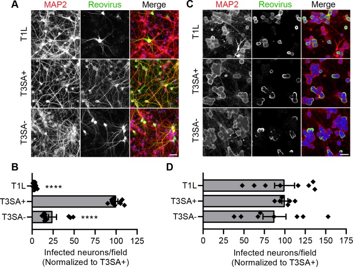
Fig 1. Serotype-dependent reovirus infection in vivo is reproduced in primary neuron cultures.
Cultured rat cortical neurons (CNs) or dorsal root ganglion neurons (DRGNs) were adsorbed at an MOI of 500 or 50 PFU per cell, respectively, with the reovirus strains shown, and cells were fixed at 24 h post-adsorption. (A, C) Neurons were immunostained using an antibody to detect a neuronal protein (MAP2) and reovirus-specific antiserum. Nuclei were stained with DAPI (blue). Representative micrographs display infected CNs (A) or DRGNs (C). Scale bars, 50 μm. (B, D) Infectivity of CNs (B) or DRGNs (D) was scored as the mean number of cells stained by reovirus antiserum per field-of-view. Bars indicate means from at least three independent experiments, each with duplicate or triplicate samples, normalized to T3SA+ infectivity. For reference, ~ 26% of CNs are infected by T3SA+ reovirus under the experimental conditions used.) Error bars indicate SEM. Individual data points are normalized averages from 8 to 12 fields-of-view from each sample. Values that differ significantly from T3SA+ by ANOVA and Dunnett's test are indicated (****, P < 0.0001).

