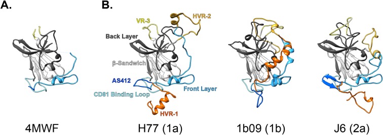
Fig 1. E2 ectodomain models.
Partial crystal structures were used as the basis for building full length models of the E2 ectodomain (defined here as residues 384–645 of the HCV polyprotein). A. PDB 4MWF partial structure of H77 E2. B. Complete E2 ectodomain models of H77, 1b09 and J6 strains; annotations denote color-coding of regions, HCV genotypes are stated in parentheses.

