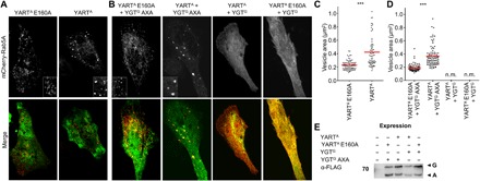
Fig. 6. Effect from YART and YGT on the Rab5A distribution in HeLa cells.

HeLa cells were transiently transfected overnight with mCherry-C1-Rab5A (top; red) together with the indicated pEGFP-C1-toxin constructs (green). Afterward, cells were washed and incubated at 21°C for an additional 3 hours. Insets show in detail Rab5A-vesicle structure. Pictures are representative of three independent experiments. Scale bar, 10 μm. (A) Effects of YARTA or YARTA E160A on the distribution of overexpressed Rab5A. (B) Effects of active YARTA and YGTG and inactive YARTA E160A and YGTG AXA on the distribution of overexpressed mCherry-C1-Rab5A. (C) Quantification by MetaMorph imaging software of the size of mCherry-C1-Rab5A vesicles in the presence of the active (YARTA) or inactive ADP-ribosyltransferase domain (YARTA E160A). Cells of view are ≥20, and n = 3. Unpaired two-sample t test was used (***P < 0.001). (D) Quantification of (B) as described in (C). n.m., not measurable. (E) For analysis of the expression of pEGFP-C1-toxin constructs, HeLa cells were lysed after transfection and expression. Proteins were analyzed by SDS-PAGE, Western blotting, and immunostaining with anti-FLAG tag antibody. G indicates GFP-YGTG and GFP-YGTG AXA; A indicates GFP-YARTA and GFP-YARTA E160A.
