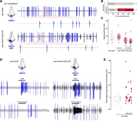
Fig. 2. Undead OSNs are functional.

(A) Representative extracellular electrophysiological traces of basal activity from neurons in an at1 sensillum of control (peb-Gal4/+) and PCD-blocked (peb-Gal4/+;UAS-p35/+) animals. Automatically detected spikes (see Materials and Methods) from the neuron expressing OR67d are shown in blue, and those of the additional, undead neuron(s) in black, as schematized in the cartoons on the left (cells fated to die are shown with dashed outlines). (B) Quantifications of the proportion of sensilla containing one neuron (gray) or two (or more) neurons (red) in control (peb-Gal4/+) and PCD-blocked (peb-Gal4/+;UAS-p35/+) animals. (C) Quantifications of the basal activity of the indicated neurons for the control and PCD-blocked genotypes. (D) Representative electrophysiological traces from at1 sensillum recordings in control (peb-Gal4/+) and PCD-blocked (peb-Gal4/+;UAS-p35/+) animals upon stimulation with a 0.5-s pulse (black horizontal bar) of the pheromone cVA [10−2 dilution (v/v) in paraffin oil] or a mix of fruit odors [butyl acetate, ethyl butyrate, 2-heptanone, hexanol, isoamyl acetate, pentyl acetate; each odor at 10−2 dilution (v/v) in paraffin oil]. Automatically detected spikes from the neuron expressing OR67d are shown in blue, and those of the undead neuron(s) in black. (E) Quantifications of odor-evoked responses to fruit odors (see Materials and Methods) in control (peb-Gal4/+) and PCD-blocked (peb-Gal4/+;UAS-p35/+) animals (n = 25 and 34, respectively).
