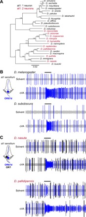
Fig. 5. Examples of naturally occurring extra neurons in at1 sensilla.

(A) Phylogeny of 26 drosophilid species, representing most of the Drosophila genus subgroups, based on the protein sequences of housekeeping loci (see Materials and Methods). Species names are colored to reflect the presence of one or two neurons in at1 sensilla. Numbers next to the tree nodes indicate the support values. The scale bar for branch length represents the number of substitutions per site. (B) Representative electrophysiological traces from recordings of at1 sensilla of the indicated drosophilid species (n = 5 per species) upon stimulation with a 0.5-s pulse (black horizontal bar) of solvent (dichloromethane) or cVA [10−2 dilution (v/v)]. A single cVA-responsive neuron (known or assumed to express OR67d orthologs) is detected (shown in blue), as schematized in the cartoon on the left. (C) Representative electrophysiological traces from recordings of at1 sensilla of the indicated drosophilid species (n = 5 per species) upon stimulation with a 0.5-s pulse (black horizontal bar) of solvent (dichloromethane) or cVA [10−2 dilution (v/v)]. Two classes of spike are detected: a cVA-responsive neuron (assumed to express OR67d orthologs) (shown in blue) and a second neuron with a larger spike amplitude, which does not respond to cVA (shown in black). The inferred sensilla organization is shown in the cartoon on the left.
