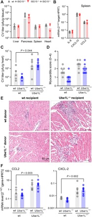
Fig. 2. Protection from CV pathology requires ISGylation in nonhematopoietic cells.

(A and B) ISG15−/− (CD45.2) mice were reconstituted with bone marrow cells from either ISG15−/− (CD45.2) or wild-type (wt, CD45.1) donors before CV infection, and mice were sacrificed 3 days after infection. (A) Infectious viral particles were quantified by plaque assay (wild-type ➔ ISG15−/−, n = 6; ISG15−/− ➔ ISG15−/−, n = 4). Data are summarized as median. (B) Splenic mRNA expression of the indicated cytokines and chemokines was determined by TaqMan qPCR. (C to F) Chimeric wild-type and Ube1L−/− mice were generated upon transfer of wild-type or Ube1L−/− bone marrow cells into lethally irradiated wild-type or Ube1L−/− recipients, respectively. Mice were infected with CV and sacrificed after 8 days (n = 7 in all four groups). (C) Infectious viral particles were quantified in heart tissue by plaque assay. Data are summarized as means ± SEM. (D) Myocarditis was scored microscopically by a blinded pathologist based on cardiac hematoxylin and eosin staining. (E) Representative histopathologic stains of heart tissue of each group are shown. (F) mRNA levels of the indicated genes in heart tissue were determined by TaqMan qPCR. Unequal variance versions of two-way ANOVA were performed, followed by a Sidak-Holm’s multiple comparison test. Data were summarized as means ± SEM if applicable.
