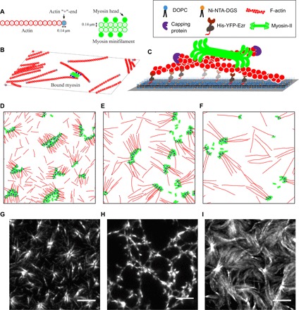
Fig. 1. Ingredients of agent-based simulations in 2D and in vitro reconstitution setup.

(A) Schematic of actin filaments (red) and myosin minifilaments (green) used in the agent-based model, with real-space dimensions indicated. One F-actin bead corresponds to 40 G-actin monomers, and one myosin bead corresponds to ∼3 to 4 heads. (B) Schematic showing a collection of F-actin and myosin minifilaments in 2D, with myosin motors bound to two actin filaments (bound myosin heads are colored blue). (C) Schematic of the in vitro reconstitution system showing the hierarchical assembly of an SLB, linker protein [His-YFP-Ezrin (HYE)], capped actin filaments, and muscle myosin-II. (D to F) Typical simulation snapshots of different aster configurations at steady state, observed in the strictly 2D simulations, as a function of increasing F-actin length la: (D) la = 2.32 μm, (E) la = 4.84 μm, and (F) la = 719 μm. The (+)-ends of the actin filaments are colored black. (G to I) Typical aster configurations at steady state observed in in vitro experiments: isolated asters, connected asters, and aster bundles, as a function of increasing F-actin length: (G) la = 2 ± 1 μm, (H) la = 3 ± 1 .5 μm, and (I) la = 8 ± 3 μm. Scale bars, 5 μm.
