. 2020 Mar 3;9:e52570. doi: 10.7554/eLife.52570

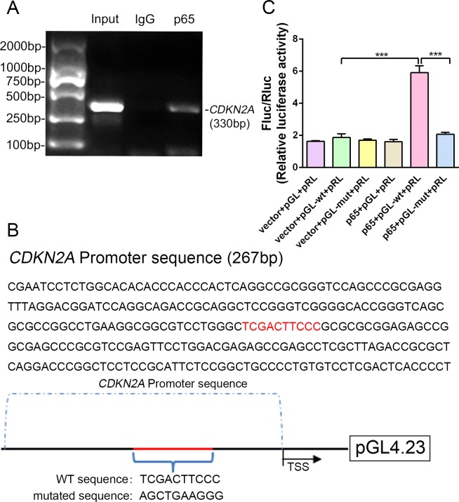
Figure 6. NF-κB-p65 bound the CDKN2A gene promoter and promoted p16 expression in human NP cells.

(A) CDKN2A promoter sequences were recovered by PCR from p65 immunoprecipitates. (B) p65‐like elements in the human CDKN2A promoter region and the mutated sequence are marked in red (upper panels). Below: structural schematic of the WT and mutant pGL4.23-p16 promoter reporter plasmids. (C) Luciferase activity driven by the CDKN2A promoter was more pronounced following NF-κB treatment. By contrast, luciferase activity that was not driven by the CDKN2A luciferase reporter decreased in the absence of NF-κB, and luciferase activity not driven by the mutant CDKN2A luciferase reporter decreased upon NF-κB treatment. Data are shown with mean ± SD (n = 3); ***p<0.001.

Figure 6—source data 1. Source data and related summary statistics for Figure 6C.

Figure 6.

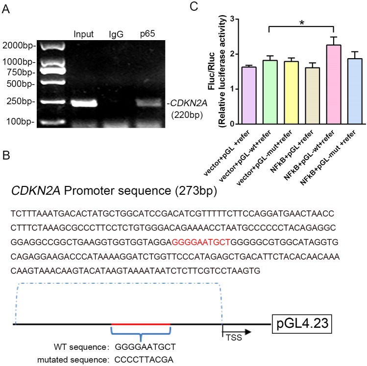
Figure 6—figure supplement 1. Another site in NF-κB-p65 that is predicted to bind the CDKN2A promoter.

Figure 6—figure supplement 1.

(A) p16 promoter sequences were recovered by PCR from p65 immunoprecipitates but not preimmune IgG immunoprecipitates. (B) p65-like elements in the human CDKN2A promoter region and the mutated sequence are highlighted in red (upper panels). Below: structural schematic of the pGL4.23-p16 promoter reporter plasmid and the mutant pGL4.23-p16 promoter reporter plasmid. (C) Luciferase activity was driven by the CDKN2A promoter, and was more dramatic following NF-κB treatment, but not by the CDKN2A luciferase reporter without NF-κB treatment. No significant difference in luciferase activity was observed following NF-κB treatment when the CDKN2A luciferase reporter was mutated. Data are presented as the mean ± SD (n = 3); *p<0.05.
Figure 6—figure supplement 1—source data 1. Source data and related summary statistics for Figure 6—figure supplement 1C.