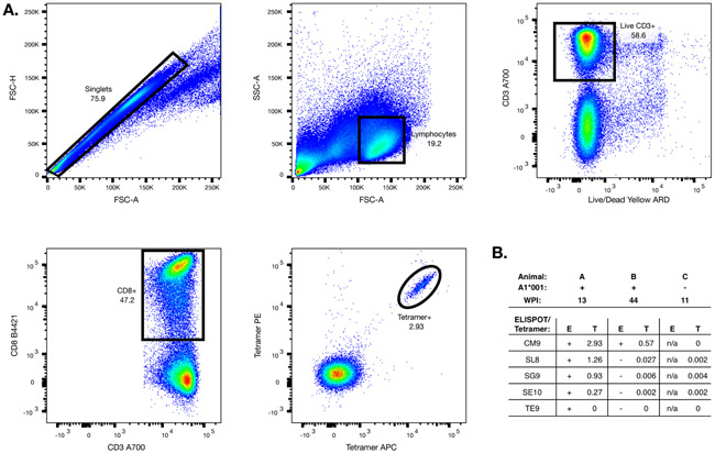
Figure 3: Tetramer analysis.
Mamu-A1*001 MHC-peptide tetramers were generated and tested for binding to PBMC from SIV-infected animals. (A) A representative sequential gating strategy for the flow cytometry data is shown. (B) Tetramer binding to PBMC from three animals was tested; shown are the ELISPOT responses to each peptide (STPESANLG, STPESANLGE, and TPESANLG are abbreviated as SG9, SE10, and TE9 respectively), and the percentage of CD8 T cells that were tetramer-positive.

