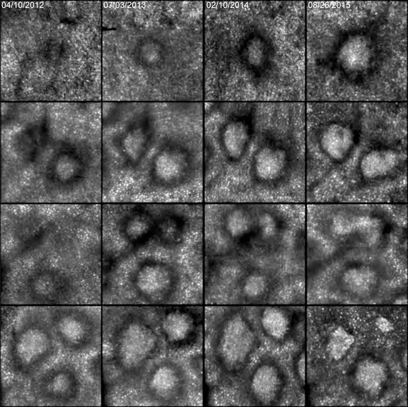
Figure 3.

Progression of individual subretinal drusenoid deposit (SDD) over 3.5 years seen in adaptive optics scanning laser ophthalmoscopy. Each row represents 1 degree (300 μm) inside the eye, imaged on the dates shown at the top of each column. Top row: one deposit grows and gains reflectivity. Second row: of two deposits, the top one starts at stage 2, grows, becomes stage 3, gaining reflectivity overall but losing reflectivity at divot-like regions. The bottom deposit starts at stage 3 with a wide annulus, and the center slightly elongates. Third row, two deposits at the top appear on slightly different time frames, then fuse. A deposit at the bottom grows to 15 months, shrinks, then grows again. Fourth row: the top left and right deposits grow and change from circular to rhomboid at 22 months, then shrink and gain reflectivity at 3.5 years. The deposit at the bottom stays at stage 3 and grows smoothly. Patient 1 in Table 1.
