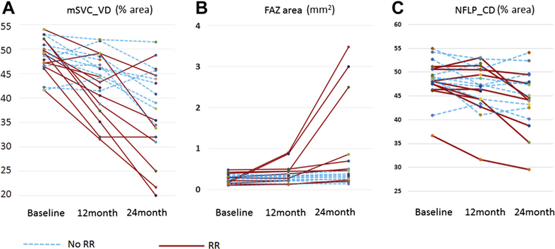
Figure 1.
Graphs showing the (A) macular superficial vascular complex vessel density (mSVC_VD), (B) foveal avascular zone (FAZ) area, and (C) peripapillary nerve fiber layer capillary density (NFLP_CD) for all patients at baseline, 12 months, and 24 months. Each line represents a single eye treated with plaque brachytherapy. Eyes with radiation retinopathy (RR) or papillopathy at 24 months are shown as solid red lines. Eyes without radiation retinopathy or papillopathy at 24 months are shown as dashed blue lines.

