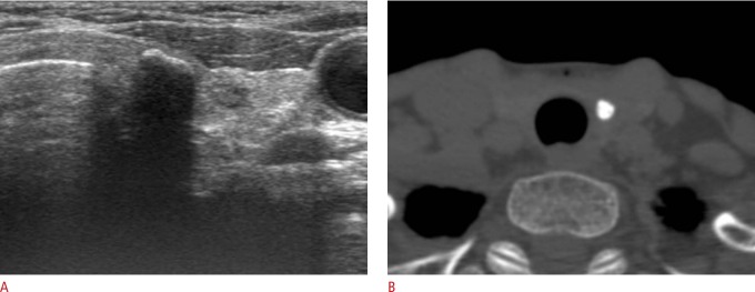
Fig. 2. Thyroid nodule with an isolated macrocalcification in a 70-year-old woman.
A. Transverse ultrasonography (US) shows a calcified nodule (6 mm) in the left lobe, and the posterior margin of the calcified nodule is not visualized due to strong posterior acoustic shadowing. B. Axial unenhanced computed tomography shows a complete compact central calcification in a case where an isolated macrocalcification was detected by US.

