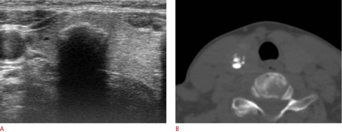
Fig. 3. Thyroid nodule with an isolated macrocalcification in a 59-year-old woman.
A. Transverse ultrasonography (US) shows a calcified nodule (12 mm) with strong posterior acoustic shadowing in the right lobe. B. Axial unenhanced computed tomography shows a partial coarse central calcification in a case where an isolated macrocalcification was detected by US.

