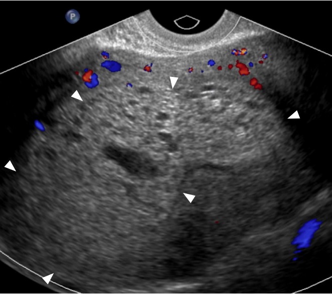
Fig. 15. Gestational trophoblastic disease: complete mole.

Transvaginal ultrasonography in a 35-year-old woman presenting with an elevated serum beta-human chorionic gonadotropin (β -hCG) level (>383,000 mIU/mL) and vaginal bleeding, shows an echogenic, heterogenous mass with minimal peripheral vascularity (arrowheads) and numerous cystic spaces. No fetal parts or myometrial invasion was identified. These findings, given the significantly elevated β-hCG value, were diagnostic of complete mole.
