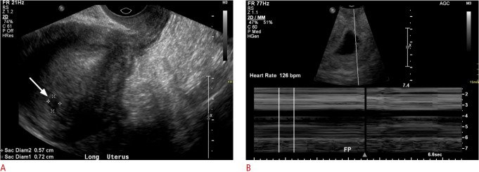
Fig. 3. A 19-year-old woman with vaginal bleeding and a positive beta-human chorionic gonadotropin test.
A. Initial transvaginal ultrasonography (TVUS) shows a vague hypoechoic collection measuring 7 mm in the uterine fundus (arrow). The morphology was not typical for an intrauterine pregnancy. B. Subsequent TVUS 2 weeks later demonstrates an intrauterine gestational sac with an embryo with heart rate of 126 bpm.

