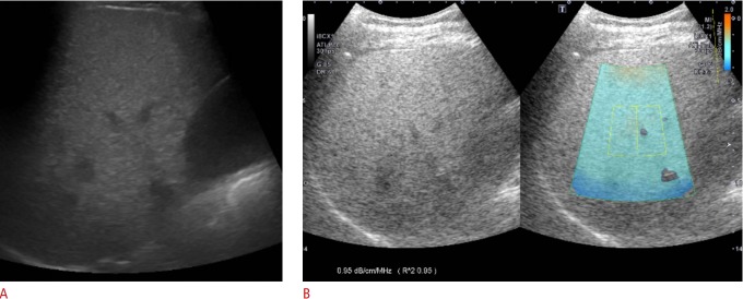
Fig. 1. Gray-scale ultrasound and attenuation imaging (ATI) in a 48-year-old man with hepatic steatosis.
A. Gray-scale ultrasound imaging shows increased liver echogenicity with obscured periportal echogenicity and preserved diaphragmatic echogenicity. Both reviewers 1 and 2 assessed the degree of steatosis as 2 (moderate). B. ATI was performed in the right lobe of the liver through an intercostal scan. The level of attenuation was color-coded and displayed in the region of interest (ROI), excluding vascular structures. The ultrasound system automatically displays the attenuation coefficient (dB/cm/MHz) and the coefficient of determination (R2 value) to optimize the accuracy of ROI placement.

