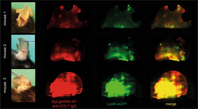
Figure 4.
Co-localization of anti-COL7 IgG and myeloid cells in the skin of mice with experimental epidermolysis bullosa acquisita demonstrates presence of myeloid cells predominantly at the anti-COL7 IgG deposits. Experimental epidermolysis bullosa acquisita was induced in LysM-eGFP+ mice (green) via repetitive i.v. injections of DyLight594 affinity purified anti-COL7 IgG (red). The distribution of the antibody deposition and localization of the myeloid cell infiltrate were determined on day 9 in whole ears using fluorescent microscopy. In all mice, red fluorescence (which corresponds to antibody deposits) co-localized with the areas with clinical lesions. Furthermore, green fluorescence (which corresponds to myeloid cells) was predominantly present at the same sites. Three representative mice from a total of 9 are shown here. Of note as a standard procedure mice are notched at weaning. Therefore, the notches appear randomly due to this procedure (mouse 1: 2 notches, mouse 2: 1 notch, mouse 3: no notch).

