Figure 5.
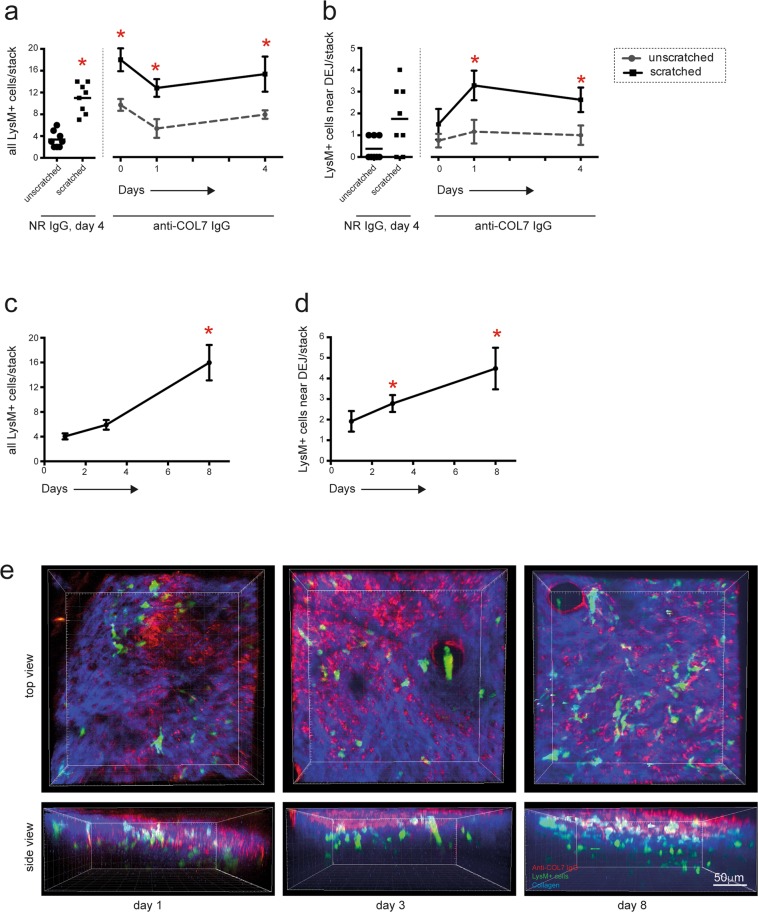
Rapid extravasation into the skin but delayed localization at the dermal-epidermal junction, of LysM-eGFP+ cells following anti-COL7 IgG injection. (a) Graph indicates the number of all LysM-eGFP+ cells identified in the dermal compartment. The bars correspond to the LysM-eGFP+ cells in the unscratched and scratched ears on day 4 after the initial normal rabbit IgG (NR IgG) i.v. injection and the lines correspond to the number of LysM-eGFP+ cells in the mice injected with anti-COL7 IgG on day 0, 1 and 4. The data are based on 6–8 evaluated stacks from at least 3 independently performed experiments [*p < 0.05, ANOVA on Ranks, followed by Dunn’s Method for comparison to reference (unscratched, normal rabbit IgG, day 4)]. (b) Here, only cells located at the dermal-epidermal junction were counted. (c) Total number of LysM-eGFP+ cells in the dermis and (d) number of LysM-eGFP+ cells located at the dermal-epidermal junction of mice i.v. injected with anti-COL7 IgG increases over the 8 day observation period. The data are based on experiments in 6 mice, and are presented as means and standard error for presentation purposes. (*p < 0.05, ANOVA on Ranks with Dunns’ Method, compared to day 1). (e) Representative stacks of the ears, generated from the images obtained by multiphoton microscopy.

