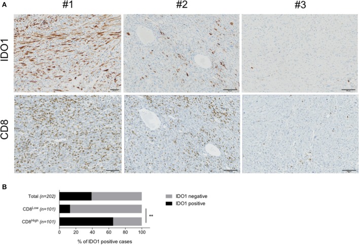
Figure 1.
IDO1 is expressed in human sarcoma and its tumor microenvironment. (A) Immunohistochemical staining for IDO1 in a cases with positive tumor cells (#1), immune cells (#2) or no positivity (#3). Stainings for CD8 in sequential sections are shown. (B) Quantification of IDO1 positivity within the total cohort (n = 203), patients harboring either high (CD8High, n = 101) or low (CD8Low, n = 101) CD8 infiltration. Data are represented as percentage. **p < 0.01.

