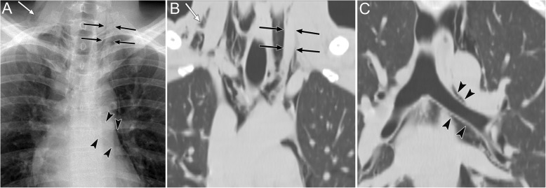
Fig. 13.
Pneumomediastinum due to chronic cough and vomiting in a 22-year-old man. a A supine radiograph shows the “tubular artery” sign (black arrows) and the “double bronchial wall” sign (black arrowheads). Subcutaneous emphysema is also evident (white arrow). b A coronal cervical CT image shows the “tubular artery” sign (black arrows). Both sides of the left internal jugular vein are visible due to pneumomediastinum. Subcutaneous emphysema is also visible (white arrow). c The coronal CT image at the carina level shows the “double bronchial wall” sign (black arrowheads). Both sides of the left bronchial wall are visible because of air in the bronchus and pneumomediastinum surrounding the wall

