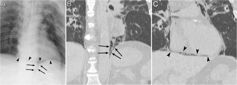
Fig. 16.
Pneumomediastinum in a 50-year-old man. The pneumomediastinum was caused by tracheostomy displacement. a A supine radiograph shows pneumomediastinum and subcutaneous emphysema. Air along the left aortic border and the medial left hemidiaphragm become V-shaped (i.e., “Naclerio’s V” sign) (arrows). The “continuous diaphragm” sign (arrowheads) is also visible. b A coronal CT image of the lung base reveals “Naclerio’s V” sign (arrows). c Another coronal CT image of the lung base reveals a “continuous diaphragm” sign (arrowheads)

