
Fig. 17.
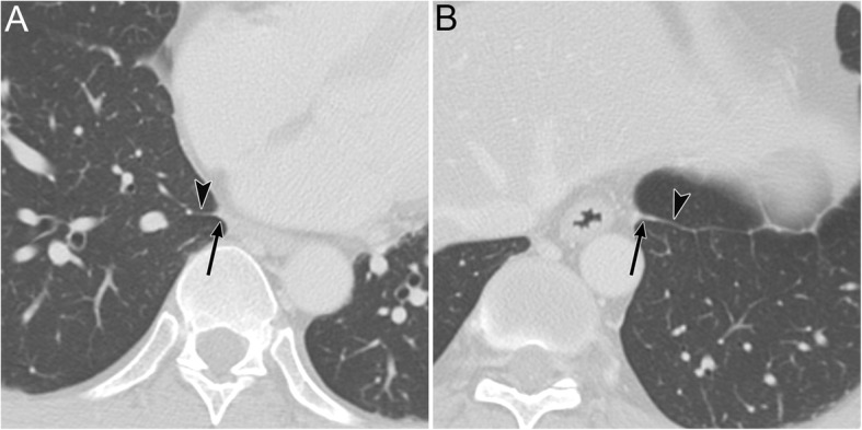
The normal inferior pulmonary ligament (IPL) on CT. A 48-year-old man with chronic obstructive pulmonary disease. a An axial CT image of the right lung at the lung base shows the right IPL (black arrow). It appears as a beak that extends laterally from the mediastinum (between the IVC and the azygos vein) with the intersublobar septum (black arrowhead). b An axial CT image of the left lung at the lung base shows the left IPL (black arrow). It appears as a beak that extends laterally from the mediastinum (along the esophagus) with the intersublobar septum (black arrowhead)