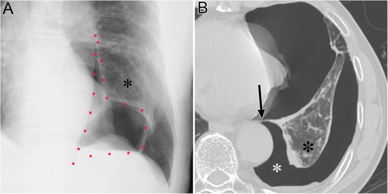
Fig. 6.

Pneumothorax in the posteromedial space of an 82-year-old man with chronic obstructive pulmonary disease. a A supine radiograph shows a lucent triangle (red dotted line) with a partially collapsed left lower lobe (black asterisk). The lucent triangle corresponds to the air collection space with its vertex in the hilum and a V-shaped base that delineates the costovertebral sulcus between the paraspinal line/descending aorta and the medial surface of the partially collapsed lower lobe. b An axial CT image of the lung bases shows pneumothorax in the posteromedial space (white asterisk). The left inferior pulmonary ligament (arrow), which is the linear structure between the mediastinum and the left lower lobe (black asterisk), are clearly visible
