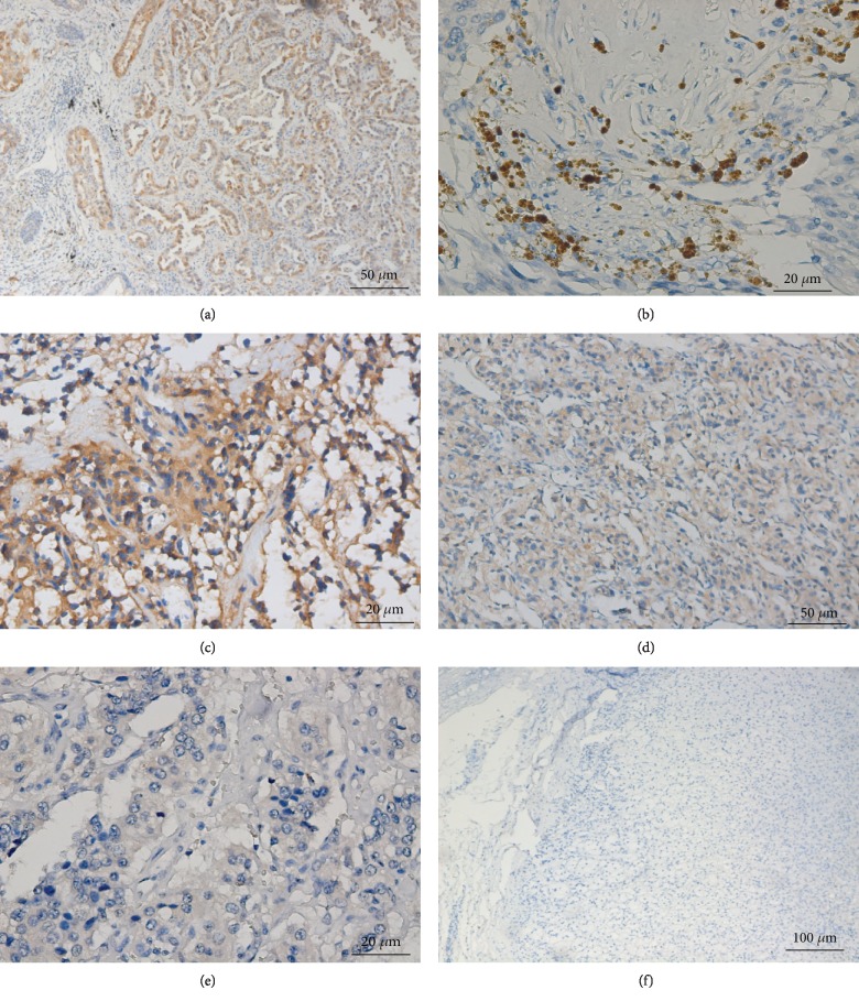
Figure 4.
Galectin-3 immunostaining in PHEO and PGL. (a) Intense staining of galectin-3 in positive control tissue (lung carcinoma) (Zeiss imager ×200). (b) Intense cytoplasm staining of galectin-3 in PPGL tumor tissue (Zeiss imager ×400). (c) Intermediate cytoplasm staining of galectin-3 in PPGL tumor tissue (Zeiss imager ×400). (d) Weak cytoplasm staining of galectin-3 in PPGL tumor tissue (Zeiss imager ×200). (e) Negative cytoplasm staining of galectin-3 in PPGL tumor tissue (Zeiss imager ×100). (f) Negative cytoplasm staining of galectin-3 in normal adrenal glands.

