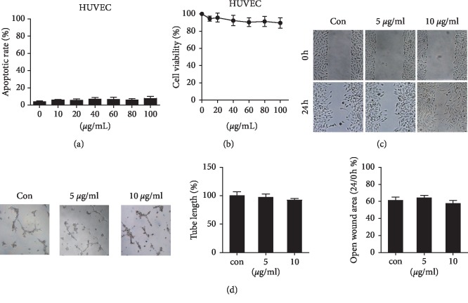
Figure 2.
At-EE did not influence HUVEC cells directly. (a–d) HUVEC cells were treated with DMSO or At-EE. (a) The apoptotic rate was assessed by flow cytometry. (b) The cell viability rate was analyzed by MTT assay. (c) Wound healing assays were done for the mobility of HUVEC cells. (d) In vitro angiogenesis evaluation was done for HUVECs treated by At-EE. The results were similar in at least three independent experiments. ∗p < 0.05. ∗∗p < 0.01.

