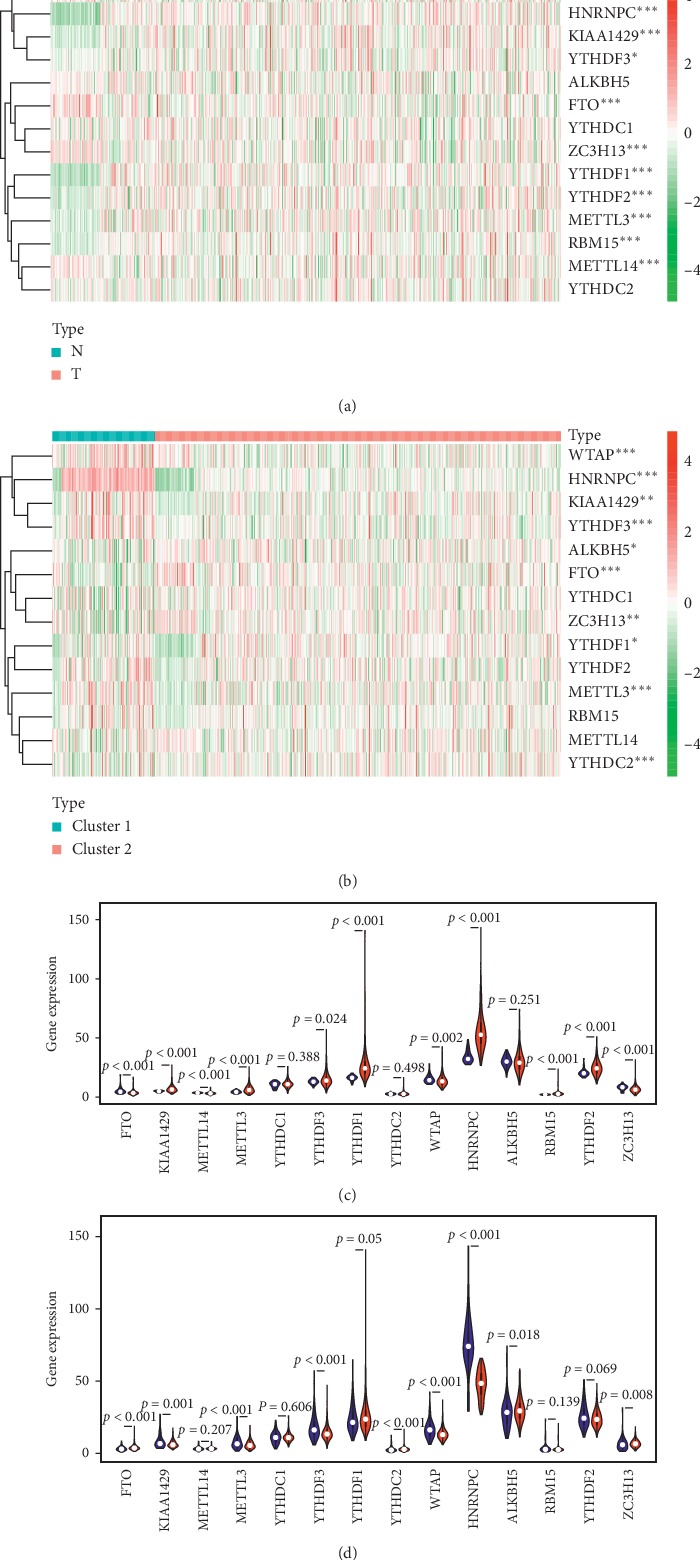
Figure 3.

The profiles of the m6A-related genes for LUAD patients. (a) The difference in gene expressions between tumor tissues and non-tumor tissues. ∗represents p < 0.05, and ∗∗∗represents p < 0.001. N = non-tumor tissues, T = tumor tissues. (b) The difference of gene expressions between cluster 1 and cluster 2. ∗represents p < 0.05, and ∗∗∗represents p < 0.001. (c) The violin plot of the m6A-related gene expressions. Blue color represents non-tumor tissues, and the red color represents tumor tissues. (d) The violin plot of the m6A-related gene expressions. Blue color represents cluster 1, and the red color represents cluster 2.
