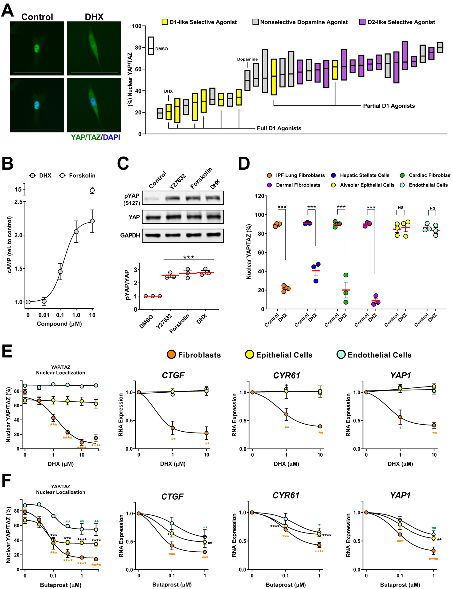
Fig. 3. DRD1 agonism selectively blocks YAP/TAZ nuclear localization in fibroblasts.

A. IPF patient-derived lung fibroblasts cells treated 2 hours prior to fixation with a library of diverse, mixed selectivity, dopaminergic agonists (10μM). N=4 biologically independent samples. Representative image: 10μM dihydrexidine (DHX). %nuclear localization of YAP/TAZ was determined using automated imaging software. Scale bar represents 100μm. Box plots represent range and mean. B. cAMP measured in IPF patient-derived fibroblasts treated for 20 minutes with the indicated concentration of DHX or forskolin (10μM). N=3 biologically independent samples. C. Total and phospho-ser127 YAP responses to the Rho-kinase inhibitor Y27632 (20μM), Forskolin (10μM), or DHX (10μM). IMR-90 lung fibroblasts N=3 independent experiments. (comparisons made using ANOVA, *** p < 0.001 vs. 0.1% DMSO vehicle control) D. YAP/TAZ nuclear localization response to DHX in fibroblasts from multiple organs: IPF patient-derived lung fibroblasts, hepatic stellate cells, human adult cardiac fibroblasts, and human dermal fibroblasts, and human lung alveolar epithelial or human pulmonary microvascular endothelial cells. N=3 biologically independent samples (comparisons made using t-test, *** p < 0.001 vs. 0.1% DMSO vehicle control). E and F. IPF derived lung fibroblasts, lung alveolar epithelial (NHAEp) and endothelial (NHMVE) cells treated with DHX (E) or with butaprost (EP2 receptor agonist, F). YAP/TAZ localization was determined after 2 hours (left), and expression of YAP/TAZ target genes at 24 hours (right). N=3 biologically independent samples (comparisons made using ANOVA, * p < 0.05, ** p < 0.01, *** p < 0.001, **** p < 0.0001 vs. the corresponding control treated cells. Orange * relate to fibroblast data, black * relate to epithelial cell data, and blue * relate to endothelial cell data).
