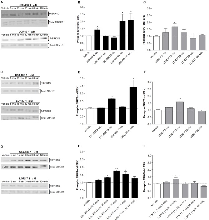
Figure 3.
U50,488- and LOR17-mediated phospho-ERK1/2 (P-ERK1/2) increase in HEK-293/hKOPr cells, U87-MG astrocytoma cells and normal human astrocytes. (A). Representative blots of HEK-293/hKOPr cells exposed to 1 µM U50,488, 1 µM LOR17 or vehicle. (B, C). Quantification of P-ERK levels in HEK-293/hKOPr cells exposed to 1 µM U50,488, 1 µM LOR17 or vehicle; data are presented as the mean ± SD of 6 independent experiments. *p < 0.05 vs vehicle (Newman-Keuls test after ANOVA). (D). Representative blots of U87-MG human astrocytoma cells exposed to 1 µM U50,488, 1 µM LOR17 or vehicle. (E, F). Quantification of P-ERK levels in U87-MG human astrocytoma cells exposed to 1 µM U50,488, 1 µM LOR17 or vehicle; data are presented as the mean ± SD of 6 independent experiments. *p < 0.05 vs vehicle (Newman-Keuls test after ANOVA). (G). Representative blots of normal human astrocytes exposed to 1 µM U50,488, 1 µM LOR17 or vehicle. (H, I). Quantification of P-ERK levels in normal human astrocytes exposed to 1 µM U50,488, 1 µM LOR17 or vehicle; data are presented as the mean ± SD of 6 independent experiments. *p < 0.05 vs vehicle; **p < 0.01 vs vehicle (Newman-Keuls test after ANOVA).

