. 2019 Oct 15;10(47):10900–10910. doi: 10.1039/c9sc04389f

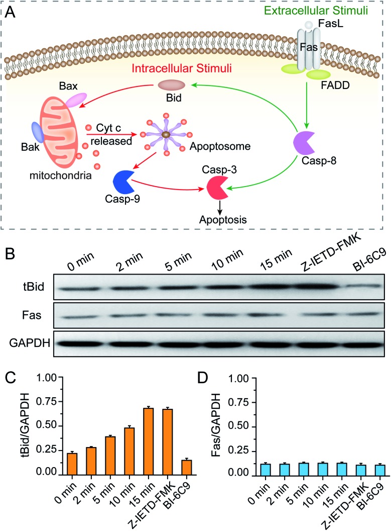
Fig. 7. (A) Illustrations of the intrinsic and extrinsic pathways of apoptosis. (B) Western blot analysis of BID and Fas activation of MCF-7 cells during the PPTT process. GAPDH served as the loading control. The expression levels of tBID (C) and Fas (D) were quantified by densitometry and normalized to the GAPDH control. Cells were incubated with mPEG/RGD/NLS-Au NSs and irradiated under an 808 nm laser for 0–15 min. Control cells were pretreated with a BID inhibitor (BI-6C9) and a caspase-8 inhibitor (Z-IETD-FMK), respectively, and subsequently incubated with the mPEG/RGD/NLS-Au NSs, and then irradiated under an 808 nm laser for 15 min.

Fig. 7