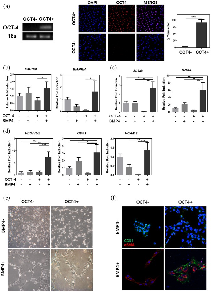
Figure 1.
Confirmation after OCT-4 transfection and BMP4 growth factor treatment. (a) Gene expression and protein expression were examined after OCT-4 transfection in HUVECs. mRNA expression levels of BMP pathway receptors (b), endothelial-mesenchymal transition markers (c), and endothelial cell markers (d) were measured after sequential treatments. (e) Bright-field images showing different morphological change depending on transfection of OCT-4 and/or treatment of BMP4. (f) Immunocytochemical staining of cells showing endothelial marker, CD31, in green and/or mesenchymal marker, α-SMA, in red. HUVECs treated or not treated with either OCT-4 or BMP4 are represented with − or + sign. The full-length agarose gels are shown in Figure S1 in Supplementary Data.
Error bars show standard deviation on the mean for n = 3: *p < 0.05; **p < 0.01; ***p < 0.001. Scale bar = 25 μm.

