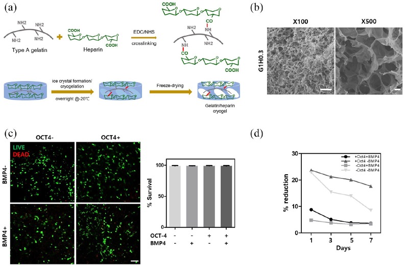
Figure 3.
Characterization of gelatin-heparin cryogel and cytotoxicity and cell metabolism measurement for cell-seeded scaffolds. (a) Schematics showing fabrication method of GH cryogels. (b) Porosity measured with SEM. (c) Live and dead images 24 h after cell seeding show scaffolds are not cytotoxic and quantification of live and dead staining. (d) Cell metabolism changing during 7 days of culture in osteogenic media. HUVECs treated or not treated with either OCT-4 or BMP4 are represented with − or + sign.
Error bars show standard deviation on the mean for n = 3: *p < 0.05; **p < 0.01; ***p < 0.001. Scale bar = 100 μm.

