Figure 4.
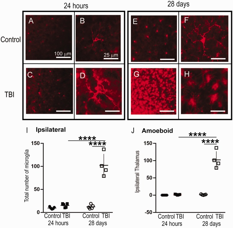
The Total Number of Microglia and Number of Amoeboid Microglia Increased in the Ipsilateral LPNT at 28 Days, but Not 24 hr, post-CCI. Panels A to H: Representative micrographs of IBA1+ cells in the ipsilateral thalamus. Panel I: There was a significant increase in the total number of IBA1+ cells in the ipsilateral LPNT of the injured brain at 28 days post-injury in comparison to both 24-hr post-injury and control. Panel J: There was a significant increase in the number of amoeboid IBA1+ cells in the ipsilateral LPNT of the injured brain at 28 days post-injury in comparison to both 24-hr post-injury and control; 24-hr cohort: TBI ipsilateral n = 5, contralateral n = 6; control ipsilateral n = 5, contralateral n = 5; 28-day cohort: TBI ipsilateral n = 3, contralateral n = 4; control ipsilateral n = 5, contralateral n = 5. Between-group comparisons were analyzed using ANOVA and if found significant, they were further analyzed using Sidak’s multiple comparison test. Statistical significance is indicated with * for p < .05, ** indicates statistical significance for p < .01, *** indicates statistical significance p < .001, and **** indicates statistical significance p < .0001. TBI = traumatic brain injury.

