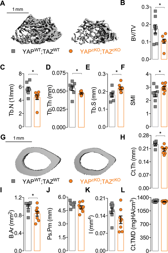
Figure 2. YAP/TAZ ablation from DMP1-expressing cells altered bone microarchitecture.
A) Representative microCT reconstructions of distal metaphysis of P84 femurs. Quantification of cancellous bone microarchitecture: (B) bone volume fraction (BV/TV), (C) trabecular thickness (Tb.Th), (D) number (Tb.N), (E) spacing (Tb.Sp), and (F) structural model index (SMI). G) Representative microCT reconstructions of mid-diaphysis cortical microarchitecture in P84 femurs. Quantification of cortical microarchitectural properties: (H) cortical thickness (Ct.Th), (I) bone area (B.Ar), (J) periosteal perimeter (Ps.Pm), (K) moment of inertia in the direction of bending (I), and (L) cortical tissue mineral density (Ct.TMD). Data are presented with individual samples in scatterplots and bars corresponding to the mean and standard error of the mean (SEM). Sample sizes, N = 8. Scale bars indicate 1 mm for microCT reconstructions.

